Technical Insight
STEM provides the answers to some difficult questions
STEM is helping to reveal information about interfaces and diffusion during device processing that is almost impossible to determine using other techniques, write Jia-Sheng Huang and Cathy Vartuli.
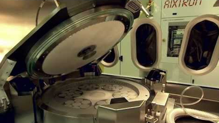
Yield improvement, cycle-time reduction and failure analysis are key elements in microelectronic chip manufacturing. To achieve high-yield and low-cost manufacturing, materials characterization is indispensable. Although there are differences in the physics and design of III-V and silicon devices, the techniques used for materials characterization are similar. Examples of the techniques routinely used for III-V and silicon materials characterization include scanning electron microscopy (SEM), energy dispersive spectroscopy (EDS), transmission electron microscopy (TEM), Auger electron spectroscopy (AES), Rutherford backscattering spectrometry (RBS), secondary ion mass spectrometry (SIMS), X-ray diffraction and atomic force microscopy (Wolf et al.).
These analytical techniques cover a range of important characterization needs. However, as device dimensions continue to shrink, the need to rapidly evaluate the elemental distributions within thin films only nanometers thick has become increasingly challenging. Recently, a dedicated scanning transmission electron microscopy (STEM) system has been introduced. The Hitachi HD-2000 thin-film evaluation system operates at 200 kV and images using secondary electrons, transmitted electrons (bright field), scattered electrons (Z-contrast or dark field) and X-rays (EDS) (Hashimoto et al.). The use of cold field-emission coupled with its lens configuration, gives a spatial resolution of 0.24 nm in bright field. The collection angle for the EDS detector is larger than a conventional TEM, giving the STEM a 2.5 times higher sensitivity for elemental analysis. The column setup allows samples too thick or rough for a traditional TEM to be evaluated, making sample preparation easier. The most powerful and unique feature of the STEM for microelectronic applications is its ability to rapidly provide high spatial resolution, high sensitivity, elemental mapping and concentration depth profiles.
The STEM technique has many applications in the characterization of III-V and silicon devices. STEM imaging can provide atomic or near-atomic spatial resolution for the study of quantum wells and dots, surfaces and interfaces, crystalline defects, grain boundary segregation and crystal tilts. Using EDS, electron energy loss spectroscopy (EELS), or Z-contrast (dark field) imaging, information about the chemical makeup of a sample or layers can be determined. Data on the sharpness of an interface, diffusion and the reaction of components can be gathered. One of the more pragmatic and alluring aspects is STEM s elemental concentration profiling for the study of interfaces in III-V or silicon materials. In the STEM concentration profile, spatial and chemical information about layers nanometers thick, as well as diffusion distance and stoichiometry, can be clearly revealed.
Strengths and limitations
STEM EDS can provide a spatially and chemically accurate elemental profile. In comparison with the various other elemental profiling techniques, STEM perhaps offers the most accurate information about the layer thickness and diffusion distance.
For AES, in situ sputtering is necessary to study the depth profile of a sample. Information from the sample is gathered from the top 2 nm, making this technique extremely useful for analyzing thin surface layers. It is difficult to determine the layer thickness in concentration profiles with high precision, due to the material dependence of the sputtering rate and the ion beam induced mixing of the sample that artificially broadens otherwise sharp interfaces. In addition, volatile elements may be preferentially removed from the material during sputtering, producing a low AES reading. Finally, the ions used for sputtering can be implanted, giving an inaccurate signal.
For RBS, the depth profile is a result of all the superimposed backscattered ion signals from the elements encountered by the probing ions within the sample. This makes the correct deciphering of a raw RBS spectrum difficult. Determining the density of the materials analyzed is important for calculating accurate concentrations. While highly sensitive to low concentrations of an element, the large beam size makes the analysis of small areas of patterned wafers impossible.
SIMS quantification of the elemental depth profile at a metal/semiconductor interface is difficult since the secondary ion yield changes drastically when sputtering from a metal to a semiconductor. While this technique is much more sensitive to trace elements than the others discussed here, the large beam size precludes its use for small devices. As the technique involves sputtering, the same problems arise in SIMS as in AES, making it difficult to accurately measure the thickness and interface quality of multiple thin layers.
SEM EDS is a rapid and relatively easy to use technique. Because it uses electrons instead of ions the problems caused by sputtering are eliminated. Depth profiles are routinely examined by viewing the cross-section of a sample, and sample preparation artifacts can be minimized. This technique is limited, however, by low spatial and chemical resolution due to the large interaction volume of the electrons generating X-rays.
TEM is able to resolve layers on the atomic scale. Measurements obtained can be calibrated to lattice images, making them highly accurate. EDS and EELS are routinely used on this tool to quantitatively determine the elements present in a sample. The lens configuration of the tool limits the solid angle of signal collected from the sample to around 0.12 mrad, constraining the sensitivity of the technique.
STEM EDS has advantages that allow it to overcome many of the challenges mentioned above. The spatial resolution of the tool is close to that of a TEM. The STEM uses a spot beam that steps across the sample to image, so EDS linescans and maps are easy to acquire. In addition, the lens configuration of this technique allows the EDS detector to be much closer to the sample, increasing the collection angle to 3 mrad, and making it 2.5 times more sensitive than a conventional TEM EDS system. While the focused ion beam (FIB) or manual polish used to make samples will create some damage to the sample surface, the interior of the sample is virtually pristine. Samples 0.5 µm and thicker are routinely used in STEM to maximize the signal and minimize the effects of sample preparation artifacts.