STM images quantum dot epitaxy inside an MOCVD chamber
Everyone working in the compound semiconductor community has heard of the atomic force microscope (AFM), a desktop instrument that can routinely analyze the surface morphology of epitaxial layers. Its importance can be gauged from its rapid adoption: still a prototype in the 1980s, less than a decade later it had been widely deployed in fabs and research centers across the globe. Before it was available, imaging of samples with a high spatial resolution had to be carried out by transmission electron microscopy (TEM), a technique that requires time consuming sample preparation and experience. Now that it is widely available, ex situ measurements of epiwafers can be carried out without any sample preparation and just by following the simple instructions provided in the manual.
Although ex situ measurements with an AFM are now routine, it would be great to use this instrument in situ and (with an atomic resolution) produce images of epitaxial processes like the growth of quantum dots. It is an aim that is still to be fulfilled. However, Bert Voigtländer and André Zinner from the Institute for Thin Films and Interfaces at the Research Centre Jülich, Germany, have used a related technique, scanning tunneling microscopy (see box "How an STM works"), to image SiGe material in an MBE reactor. Transfering this technique to an MOCVD reactor is not easy and over a decade has passed since that work before our research team at the Technical University of Berlin produced the first STM images in this harsher environment earlier this year.
Tough environmentsAn MOCVD chamber is a hot hydrogen atmosphere at near atmospheric pressure that is excellent at conducting heat and sound vibrations. Consequently, it is a poor environment for STM measurements, which require vibrations of less than 0.1 nm between the tip and sample at frequencies of above 1 Hz. This means that the sample and the STM tip must be connected together rigidly, and that the instrument must be isolated from the vibrations of the MOCVD reactor s pump and other external sources. These criteria have been met by mounting the pump on a separate plate and feeding all of the connections through sand boxes. The remaining vibrations of the susceptor-STM unit are then reduced by suspending the system with springs.
The temperature in the growth chamber also hampers STM imaging. This is because the piezoelectric tube, which provides the scanning needed to generate images, cannot operate above 150 °C due to depolarization effects and so a cooling mechanism is required. In vacuum and MBE environments, the cooling mechanism is not too demanding because the hot surfaces are small and heat transfer only occurs by radiation. In MOCVD reactor, however, the hot surfaces are much bigger and the piezoelectric tube can also be heated by conduction and convection.
We have prevented depolarization of the scan tube by adding an active cooling shield. We investigated various different designs and cooling agents before settling for a cone-shaped tube supplied with gaseous nitrogen at about 80 K (–190 °C). The latest version enables STM imaging at growth temperatures of up to 650 °C by providing a heat differential of over 400 °C across 10 mm (see figure 1a).
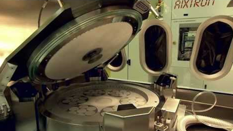
The MOCVD growth chamber also suffers from electrical noise. This is an obstacle to STM imaging, which requires maintaining a fixed tunneling current between the tip and the sample of typically 1–5 nA. Our reactor, which is a forerunner of Aixtron s AIX200 horizontal reactor, uses an inner liner quartz glass tube surrounded by a second quartz tube and provides no electrical shielding. However, we have managed to introduce some shielding by adding a thin wire mesh and we have replaced the heater s noisy power supply with a quieter DC version.
Arsenic material growth has caused us an additional problem because amorphous arsenic is a relatively good electrical conductor and can short-circuit the piezoelectric scan tube and the tunnel contact. Our solution is to balance the fluxes inside and outside the reactor s liner tube, while preventing any precursors from escaping out of the tube.
The in situ STM must not influence the epitaxial process. To ensure this, we have restricted the parts of the instrument reaching into the liner tube to the tip, the three support rods that hold the susceptor and a thermocouple for the susceptor s temperature control. The cooling shield and main STM parts are kept outside this tube and even the tip can be retracted to this region.
We tested this approach by comparing the quantum dot growth with and without the STM present. Both sets of dots were very similar and revealed a difference in growth temperature of only 5 °C, which probably resulted from variations in the thermocouple position.
Addressing all of these issues associated with in situ measurements has taken us six years and three design iterations (see figure 1 for our latest instrument). The main challenges have been designing a cooling shield and developing a compact set-up for bringing the STM tip and sample together. We have chosen a piezo motor that can travel up to 25 mm with a 4 nm resolution. The whole system is now relatively easy to maintain, can operate at reactor growth temperatures of up to 700 °C, does not influence growth and can resolve variations in surface height on an atomic scale.
Seeing quantum dots evolveOur in situ STM does not affect either the sample preparation or the growth recipes. The only change we have made is employing tertiarybutyl arsine for quantum dot growth, as this reduces arsenic contamination. Our imaging, which is undertaken after buffer growth, requires a tip with a single atom at its apex. This is produced by cooling the reactor to room-temperature before scanning at higher currents, which removes oxides and amorphous deposits from the tip and sharpens it. Depending on luck and previous treatments, this process can take from one hour to three days.
Once we have a good tip, we can heat the reactor and start to grow and image various structures, including atomic steps on GaAs epilayers (see figure 2) and InAs quantum dots (see figure 3). Monitoring quantum dot growth is not easy because the tip is colder than the surface and it picks up indium atoms, which then reduce the local growth rate. These fluctuations strongly influence the dot, which can change shape with the growth of layers just one-tenth of a monolayer thick.
To avoid these issues we have instead studied Ostwald ripening, the formation of bigger dots at the expense of smaller ones once growth has stopped. The process was observed by taking several images of the InAs quantum dots, which were grown on a GaAs substrate at 475 °C, directly after growth had ceased (see figure 3). These images reveal that the number of InAs quantum dots strongly reduces with time and those that remain increase in size. This process cannot be observed quantitatively by any other method. Ex situ methods are not suitable because the Ostwald ripening continues as the sample cools, while optical in situ methods cannot resolve the dimensions of nanometer-sized features.
Our STM development is ongoing and although we have managed to reduce preparation times from days to hours we are still spending too much time conditioning our tips. This could be avoided by using an AFM instead of an STM and we have already developed an AFM-scan head using a piezo oscillating at 3 MHz. This instrument features a laser-sharpened glass-fiber tip that can be oscillated with an amplitude of 5–10 nm and provides a height resolution of 1–3 nm.
The ultimate in situ sensor would provide chemical data and topographical information. This is possible when STMs are operated in a scanning tunneling spectroscopy mode, which measures the bandgap across the wafer s surface. With this method, local composition of ternary and quaternary alloys can be measured.
The road aheadAlthough the in situ STM holds a great promise and has already provided a unique insight into quantum dot evolution, we do not expect it to become a routine production monitoring tool. This is mainly because it is unlikely that the technique could ever be applied to growth in multi-wafer planetary and vertical reactors. In situ STM is probably also incompatible with investigations of GaN material growth because this involves reactor temperatures of at least 800 °C and probably prevent the use of piezolelectric materials. For both of these these applications we believe that engineers should instead continue to use the range of optical techniques that are already on the market.
However, we believe that a commercial STM, which could be available within five years, may have a place in the research environment. This instrument could provide a unique view of the critical steps of processes like the overgrowth of trenches, quantum dots or related problems that require a lateral resolution in the nanometer range and can fulfil the needs of a niche market.