Technical Insight
Unveiling structural anomalies in LEDs
The performance of LED light bulbs is impaired when delamination occurs within or between the parts that make up a packaged device. These deficiencies cannot be detected by widely used characterisation techniques such as X-ray and infrared microscopy, but they are exposed with acoustic imaging at very high frequencies, according to Tom Adams, a consultant to Sonoscan.
We are on the cusp of a lighting revolution. Although today’s homes are generally lit with a mix of incandescents and fluorescents, LED light bulbs are starting to make inroads, and they will dominate by the end of this decade.
If you look hard today, you’ll find that manufacturers are already offering products at eye-watering prices. These will come down, but how far and how fast suppliers drop their prices will not be the only factors determining their success – the quality and amount of light will also play a major role in the relative popularity of the various brands of LED bulb.
As LED sales rise and consumers learn more about their virtues, it will become clear how much homeowners are willing to pay for their 75 W-equivalent bulbs and how long they expect them to last. What is sure to infuriate them is if a bulb fails abruptly due to an electrical failure in its power supply or control circuitry. They will hope that the multiple LED chips in the bulb’s array will work for tens of thousands of hours. Slow dimming during that time is acceptable, while outright failure of a significant proportion of the chips will not be tolerated. A household LED bulb is generally considered to have fallen below an acceptable level of performance when its total output dips below 70 percent of its initial brightness.
Electrical failures of individual LED chips often stem from either structural anomalies in the packaging processes and materials of a semiconductor die, or in the epilayers built up on the die. These imperfections, which may have had their origin in assembly processes or in handling, include non-bonds, delaminations, cracks and voids.
In general, these weaknesses cause two types of problem: Die to overheat and fail, due to gaps at interfaces between die, die attach, substrate and heat sink that can block all the heat that needs to be removed from the assembly; and LED light output to diminish, due to the formation of unintended insulators resulting from gaps among the contact layers, blocking layers and other layers built up on the surface of the die.
Making matters worse, thermal cycling, which is inevitable when operating an LED bulb, can cause these structural gaps to expand and become more effective thermal blockers or electrical insulators.
Although LED modules are typically tested for voltage and light output, they are rarely scrutinised for internal structural defects, which can play havoc with production yield and device lifetime. Widely used techniques in the fabs, such as X-ray and infrared microscopy are unable to expose these imperfections. However, they are easily revealed with acoustic micro imaging systems based on VHF or UHF ultrasound. At Sonoscan of Elk Grove Village, IL, we have recently developed an instrument with this capability, the C-SAM system. It can be used for process control, revealing defects during production, and it can be a valuable aid for failure analysis.
Acoustic imaging may be carried out on an undiced LED wafer, or on LED arrays during assembly. In both cases, our system’s scanning transducer needs a flat surface into which to pulse the ultrasound signals. Wafers and the heat sink surface of an assembled LED are both flat; as is, at some points in production, the face of the LED. To probe an LED incorporating a lens, it may be necessary during failure analysis to grind this optical element flat to permit acoustic imaging.
The ultrasonic transducer emits pulses into the surface of the undiced wafer or individual chip, and a few millionths of a second later it reads the return echoes, resulting from reflections at interfaces. Boundaries between two solid materials tend to produce echoes of medium amplitude, while the highest amplitude echoes are returned by the solid-to-gap interfaces encountered at internal defects. The transducer carries out its pulse-echo role thousands of times a second while traversing across the sample at speeds well in excess of 1 m/s.
Imaging epiwafers...
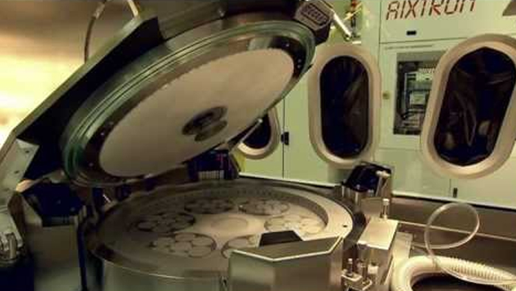
Most LED wafers feature a stack of nitride-based epitaxial layers on a substrate, typically sapphire. When imaging these wafers, our C-SAM system tends to expose delaminations and other gap-type defects, either in the sapphire substrate or the epitaxial film. These defects and anomalies may be just 5 µm or so in size, so to operate with a sufficiently high spatial resolution the transducer pulses a high frequency, such as 230 MHz. It is possible to perform manual and automated acoustic imaging with C-SAM systems. Today, the latter option is more common, with a technician placing one or more wafers on a tray, initiating the scan and examining the acoustic image to mark defective die for removal. Manual imaging makes sense when the wafer diameter is small, the value of each LED is high, and the application is critical. Typically up to sixteen 75 mm wafers can be scanned simultaneously. If the wafer is large, or if die are very small, automated inspection may be preferred.
Today, the most common LED wafer is 75 mm (3 inches), but dimensions range from 50 mm (2-inch) to 150 mm (6 inches). Eventually, 200 mm (8-inch) and 300 mm (12-inch) wafers will be used in order to gain economies of scale. This means that wafers will accommodate more die than ever before – if they are 0.3 mm x 0.3 mm in size, a common dimension for today’s LEDs, then a 300 mm wafer could yield about 700,000 chips. Such high volumes will make manual inspection more difficult and favour automated inspection.
The defects that our C-SAM system identifies are often incredibly thin delaminations. When one of the thousands of ultrasonic pulses entering the wafer each second hits the interface between solid material and a delamination, more than 99.99 percent of the ultrasound is reflected to the transducer. This incredibly high level of reflection occurs when the gap is as thin as 0.01 µm, due to the massive difference in the material properties of the solid material and the air in the delamination. This scenario produces the highest amplitude signal and identifies an internal gap. Continued scanning reveals the total area of the delamination.
With manual imaging, a technician visually identifies defective LED die in the acoustic image, so that they can be discarded from production. In comparison, with automated imaging, analysis software reports the positions of defective die via the user’s factory automation system. This allows sub-standard devices to be discarded after dicing.

Based on Sonoscan’s well-known C-SAM acoustic microscope systems and launched in 2011, the AW300 300 mm bonded wafer inspection system automates the entire inspection system from carrier attachment and wafer selection through aligning and coustic imaging to drying and sorting. Analysis software automatically measures the percentage of bonded and unbounded interface between the two wafers, and the sizes and number of voids. Accept/reject decisions are made automatically according to the user’s specific criteria
If individual devices are very small, it may be prudent to program the software to remove the eight die that are adjacent to the defective one. This makes sense when the defect is large relative to the die area, and may consequently degrade nearby die.
An example of the capability of our tool is provided in Figure 1, which shows, in very high resolution, an acoustic image of a small portion of one LED wafer. Pale grey regions indicate an absence of defects, while red features represent regions of high-amplitude reflections. These areas consist of numerous tiny gap-type defects, such as delaminations, located between various layers built up on the substrate.

Figure 1. Sonoscan’s S-CAM acoustic imaging tool reveals gap-type defects (shown in red) in a LED wafer with a sapphire base
Often LED die are small in area, and when the wafer is diced the delaminations can cause the die to fall apart. Even if these delaminations are very small, the chip can separate when scribing is used to cut up the wafers. Defective or potentially defective die must be uncovered and removed from the production process to improve the efficiency of the manufacturing process. To do this using automated analysis of images, an overlay map is employed to precisely locate each device and key each defect to a specific die for subsequent automatic removal.
...and LED assemblies
Common features of a completed LED assembly include one or more LED chips, die attach material for bonding the LED to a substrate, and attachment material for bonding the substrate to a heat sink (see Figure 2).

Figure 2. The intended pathway for thermal dissipation from a single LED is downward (red arrows) to the heat sink at the bottom. Gap-type defects, chiefly in the attachment materials, block heat and lead to early die failure. Note that the gaps shown in this figure are greatly exaggerated in their vertical dimension; gaps that are effective heat blockers and that provide high-amplitude reflection for acoustic imaging can be as thin as a fraction of a micron
The substrate on which the LED rests may be a PC board; problems with FR4 boards – a very common, flame-retardant backbone for rigid printed circuit boards – are that they have relatively high thermal resistance and transmit ultrasound poorly.
Engineers design the assembly so that it removes heat from the LED at a fast enough rate to prevent overheating – if an LED operates above its rated temperature, it will have a shorter life span. The key consideration is the total thermal resistance of the materials above and below the heat-generating LED chip.
The biggest problem – and the target of acoustic imaging – is gap-type defects. They are frequently found in: an attachment material layer or die attach layer; or between attachment material or die attach and the adjacent die, substrate or heat sink. Even extremely thin gaps can block heat transmission, leading to a hike in junction temperature and a plummeting life span for the LED.
Process engineers have the option to use our tool to inspect the topside of an LED assembly or the bottom side that features the heat sink. Scanning the latter can expose voids and other gaps in the solder between the heat sink and substrate, and between the substrate and the die. Such defects will be visible as bright features in the acoustic image. Meanwhile, scanning the top surface of the LED surface – so long as either no lens is in place, or it has been ground to create a flat scannable surface – can offer an insight into the internal features of the device.
Structural defects that are not related to heat removal can also be revealed in acoustic images. For example, images of one corner of an LED array can show features that are probably vertical cracks in the die (see Figure 3, which has a yellow arrow that marks out one of these features).

Figure 3. Acoustic imaging performed on the topside of an array of LEDs can reveal cracks, such as that highlighted by an arrow, in the sapphire chips. This array had no lens, and the depths of interest were the LEDs and their attachment to the substrate
In addition, there are some lighter-coloured features of similar structure in same area, which are probably cracks that reflect ultrasound differently, due to variations in depth and orientation; and there are four round, dark features, which are electrical connections below the die. The die themselves display a light-to-medium grey texture, indicating that they are well bonded to the substrate. There are no bright areas here that could indicate the presence of voids or disbonds (the latter are separations at an adhesive bond line in a bonded joint).
Imaging from the topside can provide valuable insights into the workings of an LED. Our acoustic imaging tool has uncovered deficiencies in a single, 0.3 x 0.3 mm die that was inspected after removing part of the polymer lens covering the die.
Pulsing ultrasound through the remaining polished lens material revealed that the square shape of the die, at centre, is somewhat distorted, due to wires attached to its upper right and lower left corners (see Figure 4).

Figure 4. After the lens has been ground flat, topside acoustic imaging of this tiny LED can reveal essentially total delamination of the polymer from the substrate
This image also shows the leads, which appear as large white features on the left and right. The red region completely surrounding the die reveals that the polymer is completely disbonded from the substrate below the die. It is possible that the disbond may already extend into the die attach material below the die, where it would reflect heat back to the die.
Expansion of the disbond under the die could also take place at a later time. This can cause individual LEDs to fail in service and to lower the efficacy of the luminaire.
Exposing these issues, which can ultimately lead to improvements in the quality of LED lighting, is one of the hallmarks of our acoustic imaging tool. As awareness of its capabilities grows, and engineers in the fabs increase their understanding of this instrument and its strengths, it will play an ever-increasing role in the solid-state lighting revolution.

Introduced in 2011, Sonoscan’s Gen6 high-end laboratory acoustic microscope is best known for its highly intuitive Sonolytics software interface and its PolyGate module. Certain samples and materials benefit from being imaged in horizontal “slices.” PolyGate permits automatic and simultaneous imaging of a sample at up to 200 different gates (horizontal “slices”)