Complementary techniques expose GaN transistor defects
Device reliability is a major challenge for GaN microelectronics. For example, AlGaN/GaN-based HFETs developed for next-generation millimeter-wave communication links and radar systems still lack the required reliability for real-life applications, even though their performance has generally reached an acceptable level. Consequently, improving reliability has been identified as a key task in DARPA s latest wide-bandgap program ("Reliability is the central issue for DARPA s triple play on GAN" Compound Semiconductor May 2005 p31).
Transistor reliability is strongly affected by self-heating effects - the increase in temperature caused by ohmic heating. Failure mechanisms are often driven by high temperatures, and greater failure rates occur at elevated temperatures. As a result, mapping a device s active temperature provides information that can be used to optimize the transistor s performance and improve its reliability.
Unfortunately, traditional thermal imaging techniques such as infrared (IR) thermography have insufficient spatial resolution to accurately measure peak device temperatures in many of today s GaN devices. For example, in AlGaN/GaN HFETs, heating occurs at the source-drain device opening, in a 0.5-1.0 μm-sized region beside the gate contact. These local temperature variations cannot be resolved with IR thermography. Instead, this technique averages temperatures over a much wider area, giving incorrect values for the device peak temperature and an underestimate of the potential risks for device failure.
Micro-Raman spectroscopy, however, can measure AlGaN/GaN HFET temperatures with sub-micrometer spatial resolution. By analyzing the phonon energies associated with lattice vibrations, the technique reveals that devices with various layouts and fabricated on different substrates can experience a temperature rise of up to 150-200 °C, even at moderate power densities.
GaN HFETs have been investigated previously with a 488 nm laser which excites the Raman spectra (see Compound Semiconductor December 2001 p26). Because this wavelength is below GaN s bandgap, the incident light produces no device heating. Measuring the changes in phonon energy produced by passing a current through a device can determine the temperature rise in the transistor s active area at the site of the micrometer-sized focused laser beam. The recorded temperatures are averages over the Raman probing depth, which is typically 1-2μ m for a confocal microscope. In practice, however, depth resolution is determined by GaN layer thickness, which is usually about 1μ m. This leads to an underestimation of the channel temperature by a few per cent.
The Raman techniqueIn principle, Raman spectroscopy can determine temperatures of up to 1200-1300 °C. The technique s spatial resolution is adequate for most device analysis considering the thermal diffusion lengths in GaN. If a higher spatial resolution is required, selected applications can use atomic force microscopy (AFM), but AFM-based thermal imaging cannot determine the device s actual temperatures unless the thermal resistance between the AFM tip and device is known. Alternative optical techniques also have their downsides. Liquid-crystal-based thermal imaging, for example, is limited by phase transition temperatures.
The Raman technique does have its own drawbacks, however. Although micro-Raman spectroscopy offers a higher spatial resolution than traditional IR thermography, it lacks the sub-second recording speed of IR thermography. Raman thermal maps are created by raster-scanning the laser beam over the device surface and dwelling for several seconds at each temperature-measurement point. Consequently, analysis is limited to relatively small areas, and so the Raman method cannot easily be applied by itself to the large ensembles of devices used for wafer screening and reliability optimization.
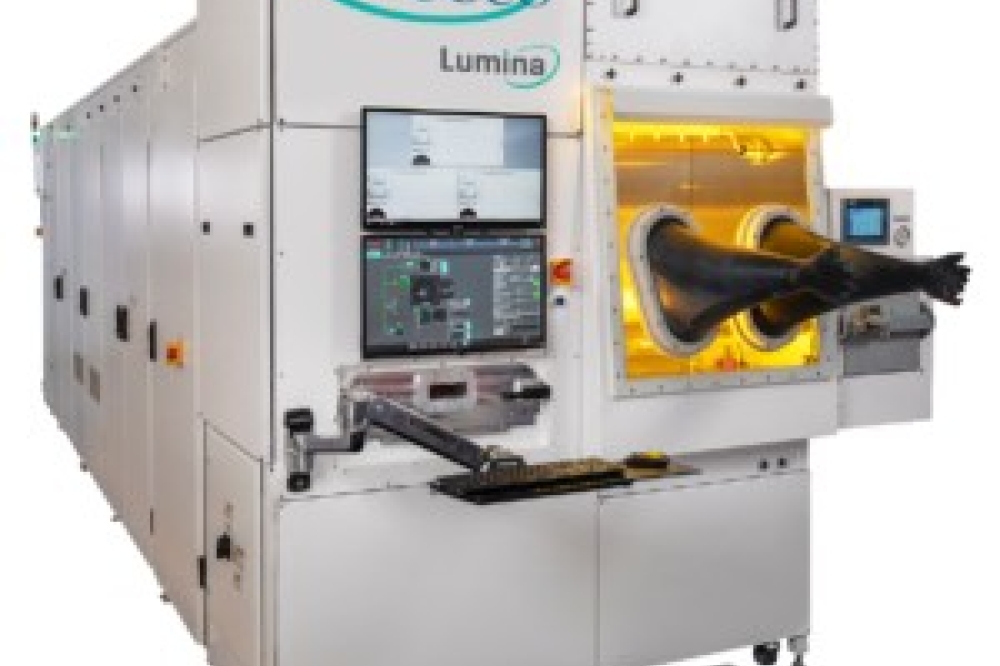
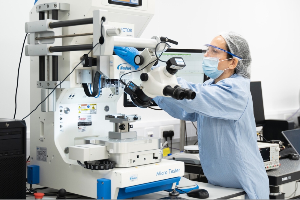
So the challenge is to deliver high-spatial-resolution temperature mapping of many transistors within a reasonable timeframe. An integrated system, containing an IR thermography instrument with an InSb detector and a Raman spectrometer, has been developed at Bristol University, UK, in partnership with Quantum Focus Instruments of San Diego, CA, and UK-based Renishaw. The collaboration is supported by UK funding agencies the Engineering and Physical Sciences Research Council (EPSRC) and the Ministry of Defence (MoD) and has recently demonstrated a prototype (figure 1).
Integrated resultsAn IR temperature map of an AlGaN/GaN HFET obtained with the integrated system, alongside a Raman temperature linescan of the device s active area, is shown in figure 2. The peak temperature rise occurs near the gate contact edge, the location of the device s highest electric field strength. As expected, the IR image provides a lower estimate of peak temperature than the Raman measurements, due to spatial averaging associated with the IR technique.
The Raman and IR measurements complement each other in several ways. The IR temperature images can quickly identify areas of interest (hotspots), but are limited to 5-10μ m resolution. The Raman spectroscopy measurements, on the other hand, deliver fast sub-micrometer resolution temperature profiles, although mapping a large area using this method would be very time consuming. Absolute temperature accuracy decreases with increasing temperature for IR measurements, while the reverse is true for the Raman technique. Using Raman spectroscopy data, temperature accuracy is about ±5 °C for temperatures in the range 100-150 °C, but this uncertainty decreases with increasing temperature, while IR images typically deliver temperature precision that is within a few per cent of the absolute temperature.
With its 1-2 °m depth resolution, and the capability to analyze phonon energies of both the GaN device layer and the underlying substrate, Raman spectroscopy can also provide three-dimensional thermal imaging (this is not possible with conventional IR thermography alone). However, unlike IR thermography, Raman measurements are unable to determine the contact temperature - unless the transistor has a flip-chip geometry, in which case measurements can be made through the substrate.
Micropipes and dislocationsIntegrating Raman spectroscopy and IR thermography produces a single instrument with the flexibility and spatial resolution needed for today s GaN microelectronics production challenges, such as wafer screening and reliability optimization. The integrated instrument has been used by UK-based QinetiQ, a partner within the EPSRC/MoD project, to optimize AlGaN/GaN HFET device performance and reliability. Raman measurements have identified transistor hotspots caused by micro-pipe and micro-crack defects in the underlying SiC substrates (figure 3). Due to their low density, which will continue to fall as the SiC substrate technology matures, these particular imperfections only tend to affect the performance of larger multi-finger devices. However, more microscopic defects such as dislocations were identified as sources for local inhomogeneous device heating, suggesting that these more common imperfections are a potentially more general source of device failure. Lateral variations in the defect density across the SiC wafer can also cause a sizable spread in device temperature that leads to variations in failure rates and reliability over a wafer (figure 4).
Optimized device packaging, such as flip-chip mounting for future "system in a package" designs, can also be developed with the integrated instrument, so long as optical access to the devices can be gained. Efforts directed at enhancing flip-chip devices, in collaboration with researchers at IMEC in Belgium, are already under way.
Although up until now the combined Raman spectroscopy and IR thermography instrument has only been applied to GaN transistors, the system is suitable for all wide-bandgap devices. And with some minor adaptations, the tool could also be used to investigate lower-bandgap silicon and GaAs devices, using the same approach to determine temperature.
The combined Raman-IR imaging tool will enable GaN manufacturers to optimize device packaging, and to address thermally related reliability and performance issues to an extent that was not previously possible. This will speed the route to real-life applications for GaN microelectronics. The integrated instrument can also be deployed to research the underlying physics of GaN devices, and help address performance issues such as the current collapse phenomenon.

