Atom probe provides evidence to question InGaN cluster theory
GaN LEDs are high-performance devices enjoying great commercial success. However, the science that underpins their operation is poorly understood, and an enthusiastic debate is currently raging within the scientific community to identify the basic mechanism for light emission from these devices.
The debate is focused on why a GaN LED, and a GaN laser for that matter, can be an efficient emitter despite its very high defect density. In other material systems, such as GaAs, these two properties could not go hand in hand because such huge dislocation densities would cause swift and catastrophic failure through non-radiative carrier recombination at dislocation cores.
With GaN, however, bright emission and high reliability is commonplace, even when dislocation densities are as high as 1010 cm–2, seven orders of magnitude higher than those found in GaAs LEDs. These very high dislocation densities arise because GaN emitters are usually grown on foreign substrates, such as SiC and sapphire, that have differing lattice constants. Although GaN bulk substrates can be produced, the process is incredibly challenging, and the crystals that are formed are currently too small and expensive for widespread commercialization.
To solve the GaN conundrum researchers throughout the community have analyzed the active region of typical devices, which contain several InGaN quantum wells approximately 2 nm thick sandwiched between GaN barriers. These experiments aimed to discover some form of microstructural feature within the quantum wells that would be responsible for the efficient excitonic emission. Such a feature could potentially ensure high radiative efficiencies by preventing migration of electron-hole pairs to non-radiative dislocation cores.
In the 1990s several researchers believed that they had found what they were looking for in transmission electron microscopy (TEM) images of the active region. Various micrographs revealed inhomogeneous strain contrast on a 2–3 nm scale in the quantum well, which was thought to result from variations in the indium content along the well. These images led to a broad consensus concerning the mechanism of light emission from these structures, which was based on exciton localization at indium-rich clusters. These clusters ensured efficient light emission because the excitons were kept away from the radiation-quenching dislocations, unless a dislocation happened to pass through an indium-rich cluster.
The trouble with TEMHowever, these TEM images were not exactly what they seemed. When Tim Smeeton from our group at the Cambridge Centre for Gallium Nitride, UK, examined some InGaN quantum wells under minimal exposure to the electron beam, he found that the TEM images actually revealed rather uniform quantum wells. It was only after further exposure that inhomogeneous strain contrast could be seen (see figure 1). This led us to believe that these "indium clusters" observed in earlier TEM studies might be nothing more than measurement artefacts, and reopened the debate concerning the nature of localization in InGaN quantum wells.
Damaging the GaN sample by introducing defects is not the only weakness of TEM. In addition, the technique in its conventional form only allows structures to be seen in projection, which means that the recorded image is a two-dimensional projection of a three-dimensional structure. Consequently, the application of a new technique that avoids electron-beam damage and delivers a fully three-dimensional set of data could be extremely beneficial.
The atom probe techniqueThe roots of such a technique were established over 50 years ago in the laboratory headed by Erwin Müller at Pennsylvania State University. On October 11, 1955 Müller and his team took the first ever atomic resolution image with the field ion microscope that he had invented. This instrument operates by ionizing the gas molecules that are above individual atoms located on the surface of a very sharp needle-shaped sample. This sample must be held at a high field, to ionize the gas, and at a very low temperature, to prevent gas molecules from moving across the surface and degrading the image s resolution.
By 1968 Müller had discovered that atoms on the sample s surface could be evaporated one at a time by superimposing a pulsed voltage onto the fixed one. Attaching a time-of-flight mass spectrometer to this instrument then allowed chemical identification of each atom. With this atomic resolution of chemical information, the atom probe was born. This instrument preceded the first atomic resolution TEM pictures by two years, and the first scanning probe microscope measurements of individual atoms by more than two decades.
Despite these great successes, the atom probe was not widely adopted. Various drawbacks plagued the first tools, such as slow collection rates, an extremely small field of view and the need for conducting samples. However, in recent years huge advances have been made in instrumentation. Fields of view of more than 100 × 100 nm are now possible, alongside detection rates exceeding a million atoms per minute. Pulsed lasers can also be used instead of pulsed voltages, enabling the atom probe to examine semiconducting and even insulating specimens.
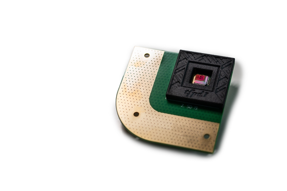
Today, sample preparation is the greatest challenge faced when using the atom probe. Samples must have a tip radius of 50 nm, and a region of interest located within a few hundred nanometers of the tip (see figure 2). For metallic samples, this is usually possible with electropolishing, but for many semiconductor samples a focused ion beam (FIB) instrument is the best tool. With this technique, gallium ion beams can mill the sample to the correct shape, with platinum or tungsten protective layers being deposited to protect the sample from ion beam damage. Some instruments also combine a scanning electron microscope with the FIB column (a FIB/SEM). This enables imaging of the sample, which can help to ensure a good tip shape, and accurate positioning of the region of interest near to the tip.
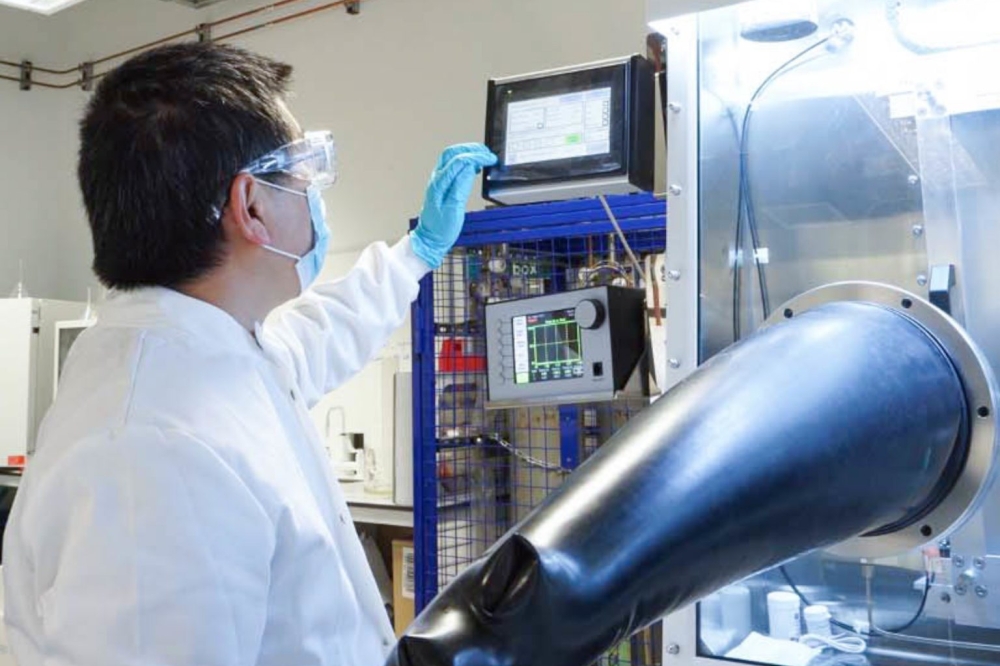
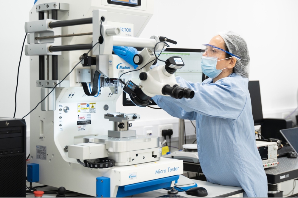
Using the latest generation of atom probes (see figure 3), such as the local electrode atom probe from US-based Imago Scientific Instruments, enables three-dimensional reconstruction of the position and chemical identity of hundreds of millions of atoms. Even elements present in very small concentrations, such as dopant atoms, can be easily detected and their distributions analyzed.
Imaging quantum wellsWe have used an atom probe to search for nanometer-scale compositional variations in GaN-based quantum-well structures, and ultimately to determine whether or not nanometer-scale indium-rich clusters are needed for bright light emission. The atom probe is an ideal technique to answer this question because it avoids the major drawbacks of the TEM, and its only serious weakness is that the sample is consumed during the measurement.
The laser-pulsed atom probe has collected data from a small volume (20 × 20 × 100 nm) of a high-quality blue-emitting InGaN/GaN multiple quantum-well structure. Reconstructing this data clearly reveals the InGaN quantum-well layers (see figure 4). Measurements of the indium content in these layers produced by atom probe agree to within 1% with compositions obtained by high-resolution X-ray diffraction, which is generally believed to be the most accurate method for determining composition.
To assess whether any clustering had occurred in the wells, we subdivided each layer into approximately 1 nm-sized boxes and calculated the composition of each box. If nanometer-scale indium-rich clusters had been present, we would have found that several of these boxes featured a much higher indium content than expected from a random alloy. In fact, we found that the distribution of the composition of the boxes fitted very well with that predicted for a random alloy. In other words, there was no evidence for nanometer-scale indium-rich clusters in this sample, despite the fact that it emitted bright light.
Now that we have unequivocally demonstrated that indium-rich clusters are not a prerequisite for bright light emission, the conundrum returns: what is the mechanism for localization? Since GaN and its alloys have a very strong piezoelectric effect, slight variations in the quantum-well thickness could be sufficient to create local energy minima that localize the carriers. The atom probe can detect nanometer-scale interface roughness, and we plan to look for this in the future. However, possible uses for the atom probe in these systems go much further, extending from basic physics studies to analysis of practical devices. Since the FIB/SEM sample preparation process is site-specific, material from failure points in LEDs could be tested to determine nanoscale changes associated with that failure. In short, this tool could provide the key to solving a whole range of problems that are fundamental to the success or failure of GaN-based light emitters.

