News Article
Using the SEM to measure crystal structures
A new procedure to measure SEM samples greatly improves its ability to measure the crystal structure of nanoparticles and extremely thin films including indium gallium nitride
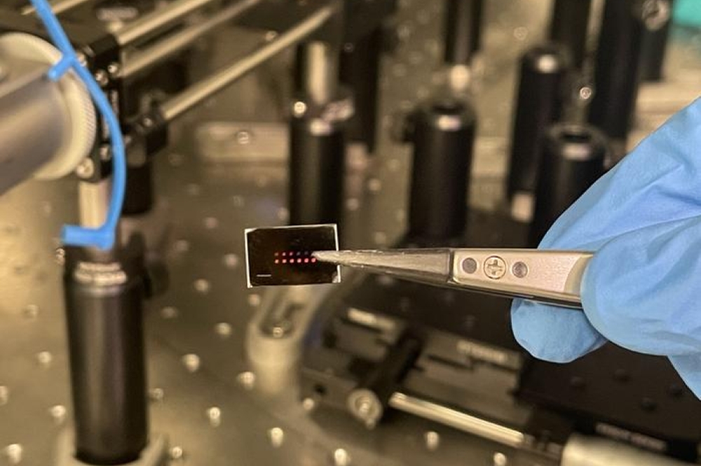
By altering sample position of materials in a scanning electron microscope (SEM), two scientists at NIST have been able to determine crystal structure of particles as small 10 nanometres (nm).
The technique, they say, should be applicable to a wide range of work, from crime scene forensics to environmental monitoring to process control in nanomanufacturing.
The technique is a new way of performing electron diffraction with an SEM. In standard SEM-based electron diffraction, the researcher analyses patterns that are formed by electrons that bounce back after striking atoms in the sample. If the sample is a crystalline material, with a regular pattern to the arrangement of atoms, these diffracted electrons form a pattern of lines that reveals the particular crystal structure or "phase" and orientation of the material.

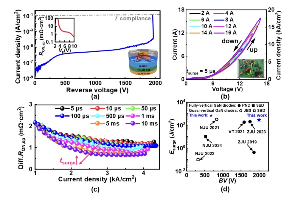
Top: Transmission electron diffraction pattern from a segment of an InGaN nanowire about 50 nm in diameter taken with an SEM using the new NIST technique clearly shows a unique pattern associated with crystal diffraction. Bottom: Same pattern but with an overlay showing the crystallographic indexing associated with the atomic structure of the material. (Credit: Geiss / NIST)
The information, say NIST's Robert Keller and Roy Geiss, can be critical. "A common example is titanium dioxide, which can exist in a couple of different crystal phases. That difference significantly affects how the material behaves chemically, how reactive it is. You need to add crystallographic identification to the chemical composition to completely characterise the material."
SEMs are often outfitted with an electron back-scatter diffraction (EBSD) detector for just this task. The problem, they say, is that below a certain size, the usual setup just doesn't work. "You can determine the crystal structure of an isolated particle down to a size of about 100 to 120 nm, but below that the crystals are so small that you're getting information about the sample holder instead." A somewhat more exotic instrument, the transmission electron microscope (TEM), does much better , but samples below about 50 nm in size show very limited diffraction patterns because the higher-powered electron beam of the TEM just blasts through them.
The novel tweak developed by Keller and Geiss combines a little of each. They moved the SEM sample holder closer to the beam source and adjust the angles so that instead of imaging electrons bouncing back from the sample, the EBSD detector is actually seeing electrons that scatter forward through the sample in a manner similar to a TEM. (They also came up with a unique method of holding samples to obtain these results.)
They have shown that their technique produces reliable crystal phase information for nanoparticles as small as 10 nm across, as well as for single crystalline grains as small as 15 nm in an ultrathin film.
Electron diffraction in an SEM, says Keller, "in general represents the only approach capable of measuring the atomic structure, defect content, or crystallographic phase of single nanoparticles. This is a critical need in cases of extremely limited sampling of unknown particles. This work pushes electron diffraction to a new frontier by providing spatial resolution that rivals that possible in a TEM, and makes it available to anyone with an SEM. And that's an ubiquitous tool in virtually all fields that require characterisation of solids."
Typical applications, the researchers say, include pinpointing ammunition sources from gunshot residue at crime scenes; determining the processing history of confiscated drugs; accurate characterisation of nanoparticles for health, safety and environmental impact studies; and optimising grain structure in high-performance electronics based on thin films and process and quality control in nanomanufacturing.
Further details of this work will be published in the paper, " Transmission EBSD from 10 nm domains in a scanning electron microscope", by R.R. Keller and R.H. Geiss in the Journal of Microscopy, 2011.
DOI: 10.1111/j.1365-2818.2011.03566.x. scheduled to appear in the March 2012 issue.