News Article
Transforming cell biology with tiny GaAs QD bioprobes
A new quantum dot device composed of gallium arsenide and light-emitting crystal, marks a new age in the study and influence of living cells. The probe could be used for real-time sensing of specific proteins within cells and be adapted to sense biomolecules such as DNA or RNA
Biological research may soon be transformed by a new class of light-emitting probes small enough to be injected into individual cells without harm to the host.
Welcome to biophotonics, a discipline at the confluence of engineering, biology and medicine in which lasers and LEDs) – are opening up new avenues in the study and influence of living cells.
Engineers at Stanford say this was the first study to demonstrate that sophisticated engineered light resonators can be inserted inside cells without damaging the cell. Even with a resonator embedded inside, a cell is able to function, migrate and reproduce as normal.
The researchers call their device a "nanobeam," because it resembles a steel I-beam with a series of round holes etched through the centre. These beams, however, are not massive, but measure only a few microns in length and just a few hundred nanometres in width and thickness.
It looks a bit like a piece from an erector set of old. The holes through the beams act like a nanoscale hall of mirrors, focusing and amplifying light at the centre of the beam in what are known as photonic cavities. These are the building blocks for nanoscale lasers and LEDs.

A photonic nanobeam inserted in a cell. Clearly visible are the etched holes through the beam as well as the sandwich-like layer structure of the beam itself. The beam structure alternates between layers of GaAs and photonic crystal containing the photon-producing quantum dots
Senior author of a paper describing the work, Jelena Vuckovic, a professor of electrical engineering, says, "Devices like the photonic cavities we have built are quite possibly the most diverse and customisable ingredients in photonics". "Applications span from fundamental physics to nanolasers and biosensors that could have profound impact on biological research."
At the cellular level, a nanobeam acts like a needle able to penetrate cell walls without injury. Once inserted, the beam emits light, yielding a remarkable array of research applications and implications.
While other groups have shown that it is possible to insert simple nanotubes and electrical nanowires into cells, nobody had yet realised such complicated optical components inside biological cells.
"We think this is quite a dramatic shift from existing applications and will enable expanded opportunities for understanding and influencing cellular biology," says the paper's first author Gary Shambat, a doctoral candidate in electrical engineering.
In this case, the studied cells came from a prostate tumour, indicating possible application for the probe in cancer research. The primary and most immediate use would be in the real-time sensing of specific proteins within the cells, but the probe could be adapted to sense any important biomolecules such as DNA or RNA.
To detect these key molecules, researchers coat the probe with certain organic molecules or antibodies that are known to attract the target proteins, just like iron to a magnet. If the desired proteins are present within the cell, they begin to accumulate on the probe and cause a slight-but-detectable shift in the wavelength of the light being emitted from the device. This shift is a positive indication that the protein is present and in what quantity.

Scanning electron microscope (SEM) image shows a nanobeam probe, including a large part of the handle tip, inserted in a typical cell.
"Let's say you have a study that is interested in whether a certain drug produces or inhibits a specific protein. Our biosensor would tell definitively if the drug was working and how well based on the colour of the light from the probe. It would be quite a powerful tool," explains Sanjiv Sam Gambhir, MD, co-author of the paper and chair of the Department of Radiology at the Stanford School of Medicine as well as director of Stanford's Canary Centre for Early Cancer Detection.
As such, embeddable nanoscale optical sensors would represent a key development in the quest for patient-specific cancer therapies - often referred to as personalised medicine - in which drugs are targeted to the patient based on efficacy.
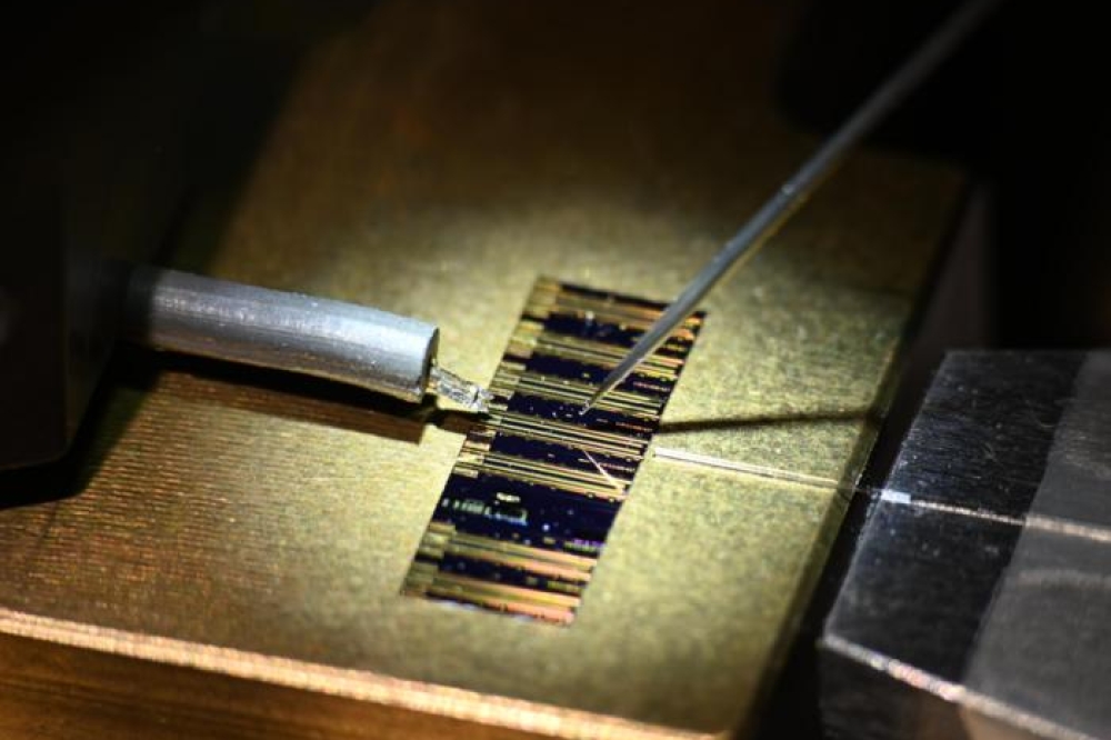
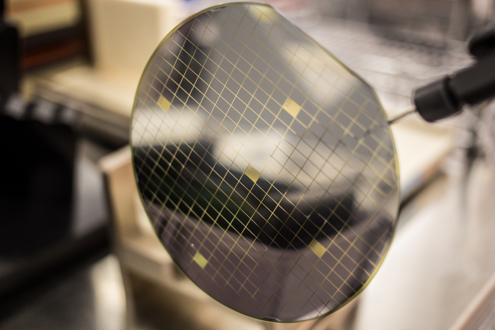
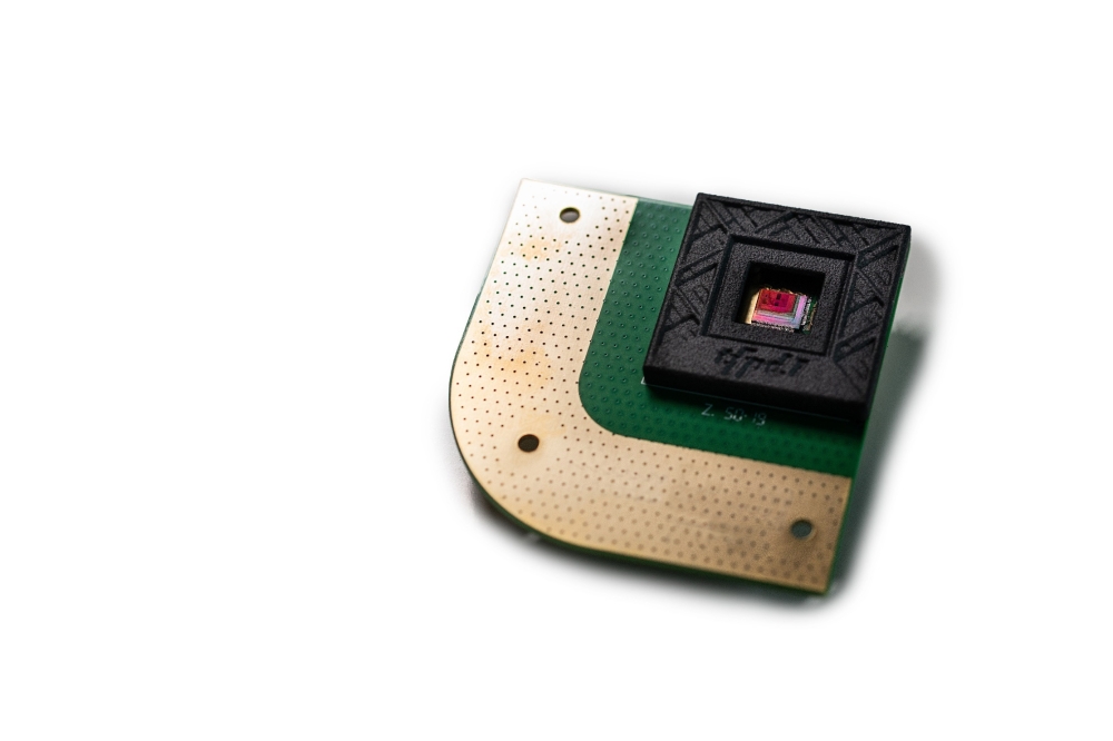
Structurally, the new device is a sandwich of extremely thin layers of GaAs alternated with similarly thin layers of light-emitting crystal, a sort of photonic fuel known as quantum dots. The structure is carved out of chips or wafers, much like sculptures are chiselled out of rock. Once sculpted, the devices remain tethered to the thick substrate.
Shambat and his fellow engineers have been working on similar optical devices for use in ultrafast, ultra-efficient computer applications where having devices immobilised on chips and wafers does not matter so much since they will ultimately be integrated with microelectronics.
For biological applications, however, the thick, heavy substrate presents a serious hurdle for interfacing with single-cells. The underlying and all-important nanocavities are locked in position on the rigid material and unable to penetrate cell walls.
Shambat's breakthrough came when he was able to peel away the photonic nanobeams, leaving the bulky wafer behind. He then glued the ultrathin photonic device to a fibre optic cable with which he steers the needle-like probe toward and into the cell.
Similarly, anticipating that GaAs could be toxic to cells, Shambat also devised a clever way to encapsulate his devices in a thin, electrically insulating coating of alumina and zirconia. The coating serves two purposes: it both protects the cell from the potentially toxic GaAs and protects the probe from degrading in the cell environment.
Once inserted in the cell, the probe emits light, which can be observed from outside. For engineers, it means that almost any current application or use of these powerful photonic devices can be translated into the previously off-limits environment of the cell interior.
In one finding that the authors describe as "stunning", they loaded their nanobeams into cells and watched as the cells grew, migrated around the research environment and reproduced. Each time a cell divided, one of the daughter cells inherited the nanobeam from the parent and the beam continued to function as expected.
This inheritability frees researchers to study living cells over long periods of time, a research advantage not possible with existing detection techniques, which require cells be either dead or fixed in place.
"Our nanoscale probes can reside in cells for long periods of time, potentially providing sensor feedback or giving control signals to the cells down the road," explains Shambat. "We tracked one cell for eight days. That's a long time for a single-cell study."
Further details of this work have been published in the paper, " Single-Cell Photonic Nanocavity Probes," by Shambat et al in Nano Letters. DOI: 10.1021/nl304602d
Funding for this study was provided by The Beckman Centre for Molecular and Genetic Medicine at Stanford, the Canary Foundation and the Centre for Cancer and Nanotechnology Excellence.
Welcome to biophotonics, a discipline at the confluence of engineering, biology and medicine in which lasers and LEDs) – are opening up new avenues in the study and influence of living cells.
Engineers at Stanford say this was the first study to demonstrate that sophisticated engineered light resonators can be inserted inside cells without damaging the cell. Even with a resonator embedded inside, a cell is able to function, migrate and reproduce as normal.
The researchers call their device a "nanobeam," because it resembles a steel I-beam with a series of round holes etched through the centre. These beams, however, are not massive, but measure only a few microns in length and just a few hundred nanometres in width and thickness.
It looks a bit like a piece from an erector set of old. The holes through the beams act like a nanoscale hall of mirrors, focusing and amplifying light at the centre of the beam in what are known as photonic cavities. These are the building blocks for nanoscale lasers and LEDs.
A photonic nanobeam inserted in a cell. Clearly visible are the etched holes through the beam as well as the sandwich-like layer structure of the beam itself. The beam structure alternates between layers of GaAs and photonic crystal containing the photon-producing quantum dots
Senior author of a paper describing the work, Jelena Vuckovic, a professor of electrical engineering, says, "Devices like the photonic cavities we have built are quite possibly the most diverse and customisable ingredients in photonics". "Applications span from fundamental physics to nanolasers and biosensors that could have profound impact on biological research."
At the cellular level, a nanobeam acts like a needle able to penetrate cell walls without injury. Once inserted, the beam emits light, yielding a remarkable array of research applications and implications.
While other groups have shown that it is possible to insert simple nanotubes and electrical nanowires into cells, nobody had yet realised such complicated optical components inside biological cells.
"We think this is quite a dramatic shift from existing applications and will enable expanded opportunities for understanding and influencing cellular biology," says the paper's first author Gary Shambat, a doctoral candidate in electrical engineering.
In this case, the studied cells came from a prostate tumour, indicating possible application for the probe in cancer research. The primary and most immediate use would be in the real-time sensing of specific proteins within the cells, but the probe could be adapted to sense any important biomolecules such as DNA or RNA.
To detect these key molecules, researchers coat the probe with certain organic molecules or antibodies that are known to attract the target proteins, just like iron to a magnet. If the desired proteins are present within the cell, they begin to accumulate on the probe and cause a slight-but-detectable shift in the wavelength of the light being emitted from the device. This shift is a positive indication that the protein is present and in what quantity.
Scanning electron microscope (SEM) image shows a nanobeam probe, including a large part of the handle tip, inserted in a typical cell.
"Let's say you have a study that is interested in whether a certain drug produces or inhibits a specific protein. Our biosensor would tell definitively if the drug was working and how well based on the colour of the light from the probe. It would be quite a powerful tool," explains Sanjiv Sam Gambhir, MD, co-author of the paper and chair of the Department of Radiology at the Stanford School of Medicine as well as director of Stanford's Canary Centre for Early Cancer Detection.
As such, embeddable nanoscale optical sensors would represent a key development in the quest for patient-specific cancer therapies - often referred to as personalised medicine - in which drugs are targeted to the patient based on efficacy.
Structurally, the new device is a sandwich of extremely thin layers of GaAs alternated with similarly thin layers of light-emitting crystal, a sort of photonic fuel known as quantum dots. The structure is carved out of chips or wafers, much like sculptures are chiselled out of rock. Once sculpted, the devices remain tethered to the thick substrate.
Shambat and his fellow engineers have been working on similar optical devices for use in ultrafast, ultra-efficient computer applications where having devices immobilised on chips and wafers does not matter so much since they will ultimately be integrated with microelectronics.
For biological applications, however, the thick, heavy substrate presents a serious hurdle for interfacing with single-cells. The underlying and all-important nanocavities are locked in position on the rigid material and unable to penetrate cell walls.
Shambat's breakthrough came when he was able to peel away the photonic nanobeams, leaving the bulky wafer behind. He then glued the ultrathin photonic device to a fibre optic cable with which he steers the needle-like probe toward and into the cell.
Similarly, anticipating that GaAs could be toxic to cells, Shambat also devised a clever way to encapsulate his devices in a thin, electrically insulating coating of alumina and zirconia. The coating serves two purposes: it both protects the cell from the potentially toxic GaAs and protects the probe from degrading in the cell environment.
Once inserted in the cell, the probe emits light, which can be observed from outside. For engineers, it means that almost any current application or use of these powerful photonic devices can be translated into the previously off-limits environment of the cell interior.
In one finding that the authors describe as "stunning", they loaded their nanobeams into cells and watched as the cells grew, migrated around the research environment and reproduced. Each time a cell divided, one of the daughter cells inherited the nanobeam from the parent and the beam continued to function as expected.
This inheritability frees researchers to study living cells over long periods of time, a research advantage not possible with existing detection techniques, which require cells be either dead or fixed in place.
"Our nanoscale probes can reside in cells for long periods of time, potentially providing sensor feedback or giving control signals to the cells down the road," explains Shambat. "We tracked one cell for eight days. That's a long time for a single-cell study."
Further details of this work have been published in the paper, " Single-Cell Photonic Nanocavity Probes," by Shambat et al in Nano Letters. DOI: 10.1021/nl304602d
Funding for this study was provided by The Beckman Centre for Molecular and Genetic Medicine at Stanford, the Canary Foundation and the Centre for Cancer and Nanotechnology Excellence.