Monolithic microLEDs eye AI
InGaN nanopyramids, produced with a single epitaxial step, promise to provide red, green and blue emission from one device.
BY AMELIE DUSSAIGNE AND BERANGERE HYOT FROM CEA-LETI AND ADRIEN MICHON AND BENJAMIN DAMILANO FROM Côte d’Azur University/CRHEA/CNRS
One of the emerging, potentially lucrative applications for III-nitride materials is augmented reality (AR). When microLEDs are made from this material systems, they can deliver a high brightness at pixel pitches down to 10 µm and less. Thanks to this accomplished performance, they are compelling candidates for high-resolution micro-displays, which lie at the heart of AR technology.
Much work has already been devoted to devising the optimum approach for manufacturing high-resolution, full-colour micro-displays. These efforts have established three leading contenders: a pick and place process, combining blue and green emitters made from III-nitrides with phosphide materials for red emission; colour conversion, realised by depositing phosphors or quantum dots on top of blue microLEDs; and monolithic integration, with the same material family providing native emission of all three primary colours.

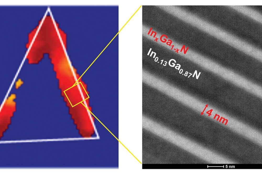
Figure 1.(a) The structure of a single InGaN nanopyramid. (b) Transmission electron microscopy image of this structure for a red-emitting nanopyramid (Gr = graphene layer).
All three solutions are progressing. Those based on picking and placing microLEDs made from different materials, and on colour conversion, are yielding some encouraging results – but that employing monolithic integration is more attractive on a number of fronts, including cost, efficiency and resolution.
For the latter, as InGaN covers the entire visible spectrum by tuning the indium content of this alloy, it is, without doubt, the material of choice for monolithic integration. However, there are issues, predominantly an external quantum efficiency that varies across the spectral range. It is high in the blue, typically 60-80 percent, but falls at longer wavelengths, dropping to 30-40 percent in the green and 10 percent or less in the red. Behind this plummet in efficiency is a degradation in the crystal quality of InGaN, with deterioration beginning at an indium content of more than 15-20 percent. To put that figure in perspective, the indium content in a blue LED is around 15 percent, that for a green LED about 25 percent, and for red LED, depending on the growth orientation, 35-40 percent.

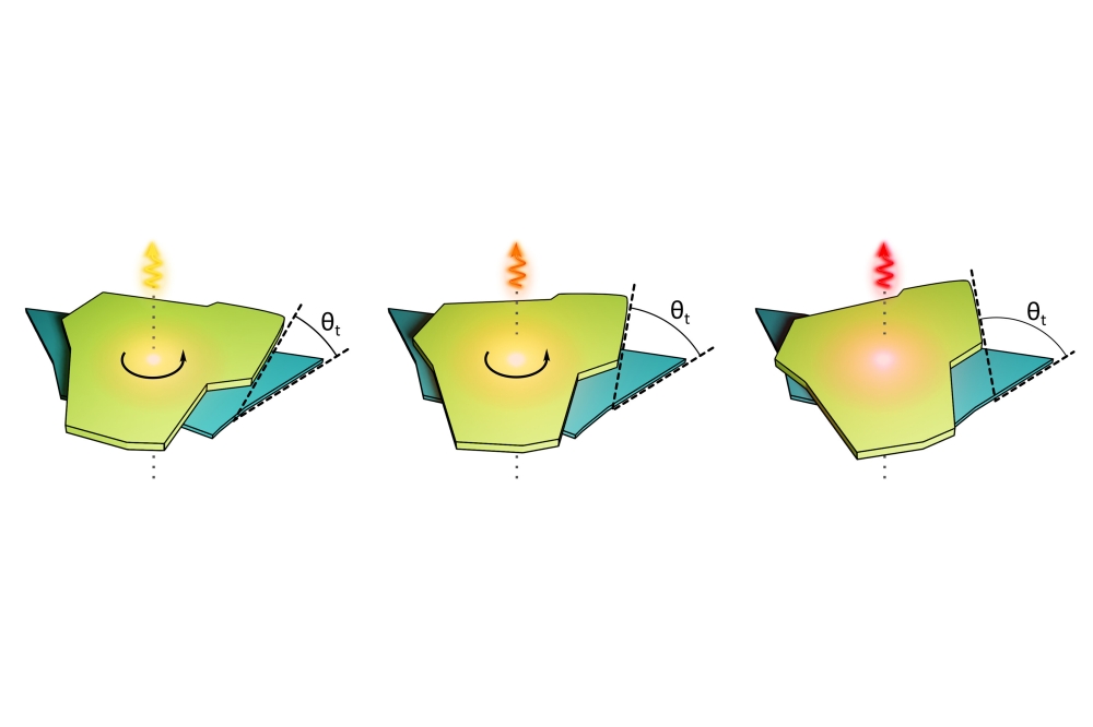
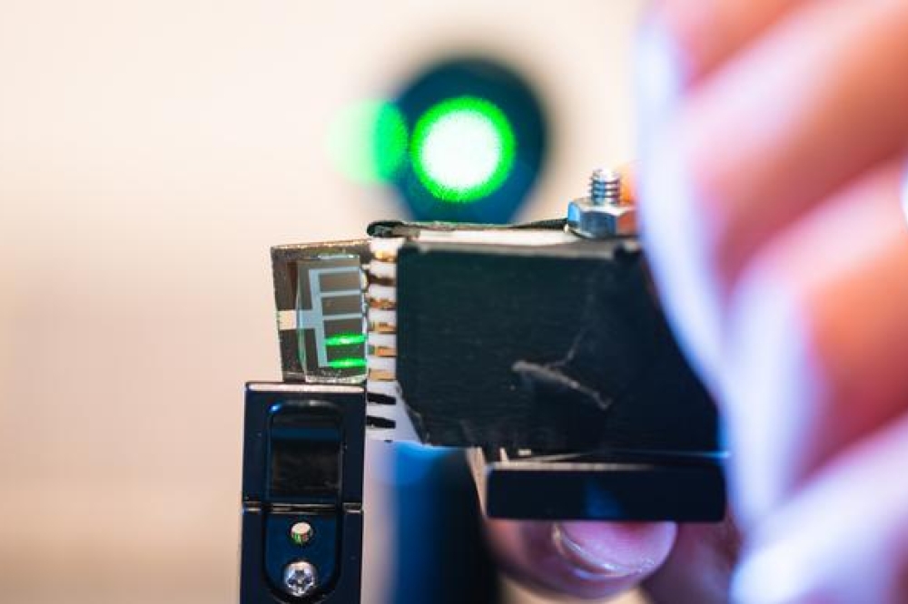
Figure 2. Photos of a 1 cm² sample under laser excitation, with emission
from blue to red depending on the position of the laser.
Due to this issue, it is clear that one of the challenges facing the development of full colour micro-displays that will employ efficient InGaN-based red, green, and blue microLEDs is the realisation of efficient InGaN-based red emission.
What’s more, if red, green and blue microLEDs are to serve in tomorrow’s AR technology, where they provide high resolution, high efficiency and reduced cost, there also needs to be a technology that enables manufacture of three-colour sub-pixels, using a monolithic integration scheme.
At CEA-Leti and CRHEA/CNRS, we are developing an approach that addresses both these matters. Our solution, involving the growth of InGaN nanopyramids through a 2D material, enables three primary colours from one epitaxial growth, using the same substrate. With this approach, we have already demonstrated regular red, green and blue quantum wells with an indium content up to 45 percent.

Figure 3. (a)-(c) Scanning electron microscopy images of the studied single nanopyramid with a lateral size of 500 nm, 750 nm and 1000 nm, respectively. (d)-(f) Cathodoluminescence (CL) average spectra acquired on the same single nanopyramid, respectively (emission from the core (dotted black line) and emission from the quantum wells (QWs) (red line)). (g)-(i) CL integrated intensity mapping for QW emission range acquired on the same single nanopyramid, respectively.
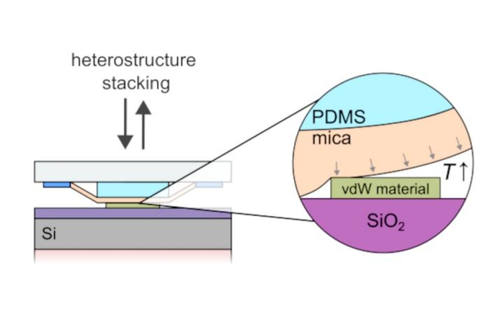
Growth of graphene
Thanks to the absence of dangling bonds on their surface, 2D materials constitute ideal masks for selective-area growth of InGaN nanopyramids. In our case, we employ a graphene mask. The graphene layer is first obtained by CVD on SiC in a hydrogen atmosphere. This method ensures self-limited growth of a single layer of graphene with a high structural quality.
If we were to attempt direct nucleation of InGaN on graphene, by either MBE or MOCVD, this would produce islands with random orientations and a lack of clear crystallographic facets. Such a foundation is incompatible with LED manufacturing. So, we locally modify graphene before InGaN growth, creating small openings around 50-100 nm in size by
H2/NH3 high-temperature treatments. These apertures allow InGaN to nucleate on the SiC substrate, beneath the graphene layer. During InGaN growth, pyramids are formed with a well-oriented crystalline structure.
InGaN nanopyramids
Following the formation of our InGaN hexagonal nanopyramids by selective-area growth, using an in situ patterned graphene monolayer on SiC as an embedded mask, we add an InGaN active zone on the nanopyramid sidewalls. This light-emitting region is composed of InGaN/InGaN multiple-quantum wells.
We have scrutinised these structures with transmission electron microscopy. Images reveal an InGaN core directly grown on SiC through small apertures in the graphene layer, and multiple quantum wells on the sidewalls of the pyramids (see Figure 1).
When we use transmission electron microscopy to provide a wider view, we see that the InGaN nuclei appear preferentially organised along specific directions, corresponding to step edges of the SiC substrate. Since the graphene layer on these edges is more defective than on the terraces, the conditions are favourable for nucleation of the InGaN nanopyramids on the SiC substrate.

Figure 4. High-resolution transmission electron microscopy images performed on a single nanopyramid emitting in the (a) green, (b) orange, and (c) red range. Corresponding CL average spectra of the quantum well areas recorded on the same transmission electron microscopy lamellas.
Red, green and blue
Under UV laser excitation, we observe emission spanning the blue to the red on different areas of our 1 cm² sample (see Figure 2). Red emission is centred at 645 nm at room temperature.
Crucially, different emission wavelengths are realised with a single epitaxial growth. This demonstrates it’s possible to obtain red, green and blue emission via monolithic integration, using just one epitaxial growth.
Cathodoluminescence mappings on single nanopyramids (see Figure 3) reveals that emission wavelengths are correlated to the lateral sizes of the nanopyramids: cyan emission for small nanopyramids, around 500 nm in size; green-yellow emission for medium-sized nanopyramids, around 750 nm in size; and amber-red emission for larger pyramids, with dimensions of about 1 µm. We have also observed that emission is homogeneous on the facets and edges of the nanopyramids, with a higher intensity at the edges, due to superior light-extraction efficiency.
Another noteworthy observation is that differences in the nanopyramid lateral size are related to differences in nanopyramid densities – a small lateral size is accompanied by a high density, and a large lateral size occurs alongside a low density. This makes sense, given that during epitaxial growth, material is distributed in each area according to the available sidewall total surface, so there will be variations in the thicknesses and indium content in the InGaN wells and barriers of the active region. Note that at this stage, the graphene mask presents a self-organized hole pattern created during the initial in-situ annealing step.
To assess the crystalline quality and optical properties of different quantum-well regions, we extracted three transmission-electron-microscopy lamellae, taken from three different areas of the sample. On each lamella – they had lateral sizes of 500 nm, 750 nm and 1000 nm – we conducted transmission electron microscopy and cathodoluminescence characterisation (see Figure 4). Results revealed that the wells are regular, with an average width from 3.5 nm to 4 nm, and homogeneous in terms of chemical contrast, which is a sign of a homogeneous indium distribution in the InGaN alloy.
To gain greater insight into our red-emitting nanopyramids, we obtained transmission electron microscopy images of the whole nanopyramid, and also zoomed in on the quantum-well area (see Figure 5).
In addition, we conducted cathodoluminescence wavelength mapping, finding red emission homogeneously distributed along the sidewalls (see Figure 5 (b)); and obtained an electron dispersive X-ray spectroscopy profile, taken from the core of the nanopyramid through the quantum wells. The latter revealed an indium content of 13 percent in the core, and 42-45 percent in the wells – that’s a record indium content in the quantum-well area, realised in conjunction with homogeneity in this region.

Figure 5. (a) Transmission electron microscopy (TEM) images of the red
emitting whole nanopyramid and of the quantum well (QW) area. (b)
Cathodoluminescence wavelength mapping on the same TEM lamella. (c)
Electron dispersive X-ray spectroscopy profile, starting from the core
through the QW area.
According to strain mapping, the core of our nano-pyramids is at least nearly relaxed, from base to apex. The implication is that a relaxed InGaN pseudo-substrate, in our case the pyramid core with a 13 percent indium content, helps enhance the indium incorporation rate in the InGaN alloy without degrading crystal quality. The advantage of the graphene mask, which benefits from the presence of the Van der Waals gap, is that it provides almost free-standing nanopyramids – they are linked to the SiC substrate by just a hole that’s 80-100 nm in diameter, with a height of 2 monolayers.
Our efforts show that it’s possible to produce a light-emitting structure on the same substrate, with just a single growth run, that provides red, green and blue emission. Additional key findings are that our approach can produce relaxed InGaN pseudo-substrates that enhance indium incorporation in the InGaN alloy beyond the theoretical limit of 25 percent when grown on GaN, a feat that’s accomplished without degrading crystalline quality.
One of our next goals is to define an organized hole pattern in the graphene mask, so that we can form red, green and blue sub-pixels next to each other, by playing on the nanopyramid diameters and densities.
We also plan to advance our process integration of our nanoLEDs, by transfer of the nanopyramids to a conductive substrate. The electrical contacts will be taken from the back side for the n contact, and from the nanopyramid sidewalls for the p contact.
To conclude, our combination of a graphene mask and a full InGaN structure is an attractive solution to realising red, green and blue emission from a single epitaxial growth while maintaining high crystalline quality in the active area. This is even realised with the red-emitting structure, a breakthrough that promises high efficiency for all three InGaN-based primary colours.
Part of this research was supported by the “Recherche Technologique de Base” and “France 2030 - ANR-22-PEEL-0014” programmes of the French National Research Agency (ANR).
The authors wish to thank the contributions provided by C. Paillet, N. Rochat, D. Cooper, and A. Grenier from CEA-Leti, and S. Vezian from CRHEA/CNRS. Note that C. Paillet is now at Aixtron.